41 human eye with labels
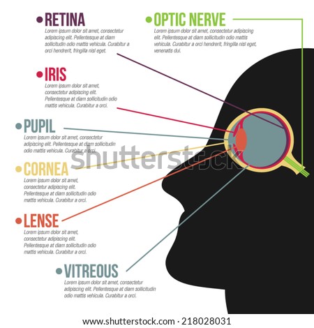
File:Diagram of human eye without labels.svg - Wikimedia Commons File:Diagram of human eye without labels.svg. Size of this PNG preview of this SVG file: 410 × 430 pixels. Other resolutions: 229 × 240 pixels | 458 × 480 pixels | 732 × 768 pixels | 976 × 1,024 pixels | 1,953 × 2,048 pixels. Labeled Eye Diagram | Science Trends What you want to interpret as a major part of the human eye is somewhat up to the individual, but in general there are seven parts of the human eye: the cornea, the pupil, the iris, the lens, the vitreous humor, the retina, and the sclera. Let's take a closer look at each of these components individually. The Cornea
Eye Diagram With Labels and detailed description - BYJUS Iris is the coloured part of the eye and controls the amount of light entering the eye by regulating the size of the pupil. The lens is located just behind the iris. Its function is to focus the light on the retina. The optic nerve transmits electrical signals from the retina to the brain. Pupil is the opening at the centre of the iris.
Human eye with labels
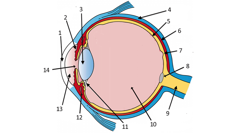
43 diagram of the human eye without labels Label Parts of the Human Eye - University of Dayton Label Parts of the Human Eye. Select One Anterior Chamber Ciliary Body Cornea Fibrous Tunic Iris Lateral Rectus Muscle Lens Medial Rectus Muscle Optic Disk Optic Nerve Pupil Retina Vascular Tunic Vitreous Nerve. Human Eye Anatomy - Parts of the Eye ... Human Eye Diagram, How The Eye Work -15 Amazing Facts of Eye The shark has even been used in human eye surgery! FACT 4 The length of our eyes are about 1 inch across and weigh about 0.25 ounce. FACT 5 Our eyeballs stay the same size forever but our nose and ears continue to grow. FACT 6 Eyes are the second most complex organ after the brain. Human Eye Anatomy Pictures, Images and Stock Photos Anatomy of human eye and descriptions. Parts of the eye, labeled vector illustration diagram How eye work medical illustration, eye - brain diagram, eye... human blue eye extreme macro Eye anatomy. Rod cells and cone cells. Anatomy of human eye hand draw vintage clip art isolated on... eyeball Human eye anatomy Human eye anatomy vector design
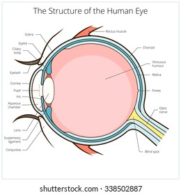
Human eye with labels. Human Eye Diagram Without Labels - solved label this diagram of a human ... Human Eye Diagram Without Labels. Published by Carol; Monday, April 25, 2022 Eye anatomy: A closer look at the parts of the eye In a number of ways, the human eye works much like a digital camera: Light is focused primarily by the cornea — the clear front surface of the eye, which acts like a camera lens. The iris of the eye functions like the diaphragm of a camera, controlling the amount of light reaching the back of the eye by automatically adjusting the size of the ... Draw And Label The Structure Of The Human Eye A human eye is roughly 23 cm in diameter and is almost a spherical ball filled with some fluid. It is the outer covering a protective tough white layer called the sclera white part of the eye. Cornea Iris Pupil Ciliary muscles Eye lens retina and optical nerves which are labelled in the diagram below. Labelled Diagram of Human Eye, Explanation and Function - VEDANTU The human eye is a part of the sensory nervous system. Labeled Diagram of Human Eye The eyes of all mammals consist of a non-image-forming photosensitive ganglion within the retina which receives light, adjusts the dimensions of the pupil, regulates the availability of melatonin hormones, and also entertains the body clock.
PDF Eye Anatomy Handout - National Eye Institute of light entering the eye. Lens: The lens is a clear part of the eye behind the iris that helps to focus light, or an image, on the retina. Macula: The macula is the small, sensitive area of the retina that gives central vision. It is located in the center of the retina. Optic nerve: The optic nerve is the largest sensory nerve of the eye. ellenjmchenry.com › uploads › 2016CUT-AND-ASSEMBLE PAPER EYE MODEL • thin permanent marker for a number labels on plastic parts (such as a very thin point Sharpie) Assembly: 1) After copying pattern pages onto card stock, cut out all parts. On the background page that says THE HUMAN EYE, cut away the black rectangles and trim the triangles at the bottom, as shown in picture above. The Eye - Science Quiz - Seterra Our eyes are highly specialized organs that take in the light reflected off our surroundings and transform it into electrical impulses to send to the brain. The anatomy of the eye is fascinating, and this quiz game will help you memorize the 12 parts of the eye with ease. academic.udayton.edu › psy323 › labelsLabel Parts of the Human Eye - University of Dayton Parts of the Eye Select the correct label for each part of the eye. The image is taken from above the left eye. Click on the Score button to see how you did. Incorrect answers will be marked in red.
› photos › male-human-anatomyMale Human Anatomy Diagram Pictures, Images and Stock Photos Human anatomy, back injury or disease, medical concepts. Three main curvatures of the spine disorders or deformities on male body: lordosis, kyphosis and scoliosis 3D rendering illustration. Human anatomy, back injury or disease, medical concepts. male human anatomy diagram stock pictures, royalty-free photos & images PDF Parts of the Eye Eye Diagram Handout Author: National Eye Health Education Program of the National Eye Institute, National Institutes of Health Subject: Handout illustrating parts of the eye Keywords: parts of the eye, eye diagram, vitreous gel, iris, cornea, pupil, lens, optic nerve, macula, retina Created Date: 12/16/2011 12:39:09 PM en.wikipedia.org › wiki › Human_eyeHuman eye - Wikipedia The human eye is a sensory organ, part of the sensory nervous system, that reacts to visible light and allows us to use visual information for various purposes including seeing things, keeping our balance, and maintaining circadian rhythm . The eye can be considered as a living optical device. tinybop.com › apps › the-human-bodyThe Human Body educational app for curious kids by Tinybop ... Interactive labels in over 50 languages, including Chinese, Russian, German, French, Spanish, Japanese — and more! Meet the artist Designer and tinker Kelli Anderson created more than 200 drawings for The Human Body .
Basic Humans Eye Anatomy Vector Infographic Stock Vector (Royalty Free) 218028031 - Shutterstock
Human Eyes Stickers | Zazzle Decorate water bottles, envelopes, clothing and more with Human Eyes stickers & labels from Zazzle! Choose from thousands of designs or create your own today! Decorate water bottles, envelopes, clothing and more with Human Eyes stickers & labels from Zazzle! ... Human Eye Crying Tears Flowing Drawing Square Sticker. $8.15. 40% Off with code ...

Eye Anatomy: 16 Parts of the Eye & Their Functions The following are parts of the human eyes and their functions: 1. Conjunctiva The conjunctiva is the membrane covering the sclera (white portion of your eye). The conjunctiva also covers the interior of your eyelids. Conjunctivitis, often known as pink eye, occurs when this thin membrane becomes inflamed or swollen.

33 Human Eye With Label - Labels Design Ideas 2020
en.wikipedia.org › wiki › Human-readable_mediumHuman-readable medium - Wikipedia In computing, human-readable data is often encoded as ASCII or Unicode text, rather than as binary data. In most contexts, the alternative to a human-readable representation is a machine-readable format or medium of data primarily designed for reading by electronic, mechanical or optical devices, or computers.
Easy Human Eye Diagram With Labels - Diagram Media
The Human Eye Label Diagram | Quizlet Start studying The Human Eye Label. Learn vocabulary, terms, and more with flashcards, games, and other study tools.

WMU Psychology Department: Lisa Baker
Labelling the eye — Science Learning Hub In this interactive, you can label parts of the human eye. Use your mouse or finger to hover over a box to highlight the part to be named. Drag and drop the text labels onto the boxes next to the eye diagram If you want to redo an answer, click on the box and the answer will go back to the top so you can move it to another box.

Human Anatomy Lab: Muscles of the Arm
Labelling the eye — Science Learning Hub Labelling the eye Add to collection The human eye contains structures that allow it to perceive light, movement and colour differences. In this activity, students use online or paper resources to identity and label the main parts of the human eye. By the end of this activity, students should be able to: identify the main parts of the human eye

World All Animals: Hippopotamuses New profile and Images
Human eye diagram Images, Stock Photos & Vectors - Shutterstock Find Human eye diagram stock images in HD and millions of other royalty-free stock photos, illustrations and vectors in the Shutterstock collection. Thousands of new, high-quality pictures added every day.

Anatomy of the Eye
Structure and Functions of Human Eye with labelled Diagram The human eye is a roughly spherical organ, responsible for perceiving visual stimuli. It is enclosed within the eye sockets in the skull and is anchored down by muscles within the sockets. Anatomically, the eye comprises two components fused into one; hence, it does not possess a perfect spherical shape.

Discovering Something New -- ongoing learning: How the eye works
The Eyes (Human Anatomy): Diagram, Optic Nerve, Iris, Cornea, Pupil, & More Your eye is a slightly asymmetrical globe, about an inch in diameter. The front part (what you see in the mirror) includes: Iris: the colored part. Cornea: a clear dome over the iris. Pupil: the ...

302 Found
› Can-C-Drops-Milliliter-LiquidAmazon.com: Can-C Eye Drops 5ml Liquid (2 in 1 Pack) Can C ... This item Can-C Eye Drops 5ml Liquid (2 in 1 Pack) Can C Cataract Eye Drops N-Acetylcarnosine, Human and Animal Eye, Cataract Eye Drops for Dog - Gift Set with Boxiti Wipe HealthCareAisle Eye Allergy Itch Relief - Olopatadine Hydrochloride Ophthalmic Solution USP, 0.2% – 2.5mL – Eye Allergy Drops

Fedex Driver Resume Samples | QwikResume
The Human Eye (Eyeball) Diagram, Parts and Pictures The eyeball is a round gelatinous organ that contains the actual optical apparatus. It is approximately 25 mm in diameter and sits snugly in the orbit where six muscles control its movement. The eyeball has three layers, each of which has several important structures that are essential for the sense of vision. Wall of the Eyeball
31 Label The Eye Quiz - Best Labeling Ideas
39 diagram of the human eye without labels Draw a labeled diagram of human eye. Write the functions ... Cornea of the eye is the first sight where convergence of light rays takes place. Iris is that part of the eye which controls the amount of light entering the eye through the pupil. Pupil is a type of small hole through which light enters the eye.

picture front of the eye without labels clipart - Clipground
Human eye Label the structure below to review the | Chegg.com Transcribed image text: 4 Human eye 2.94 points Label the structure below to review the anatomy of the human eye. Place your cursor on the boxes for the function of each structure. fovea fovea eBook sclera retina artery Print retina sclera References lens optic nerve pupil vein lens optic nerve vein ciliary muscle ciliary muscle iris artery cornea pupil cornea iris

More Then 50 Best Tattoo Designs 2014 For Men ~ 8FACT
Human Eye Anatomy Pictures, Images and Stock Photos Anatomy of human eye and descriptions. Parts of the eye, labeled vector illustration diagram How eye work medical illustration, eye - brain diagram, eye... human blue eye extreme macro Eye anatomy. Rod cells and cone cells. Anatomy of human eye hand draw vintage clip art isolated on... eyeball Human eye anatomy Human eye anatomy vector design

639 best images about Medical Terminology Help Learning on Pinterest
Human Eye Diagram, How The Eye Work -15 Amazing Facts of Eye The shark has even been used in human eye surgery! FACT 4 The length of our eyes are about 1 inch across and weigh about 0.25 ounce. FACT 5 Our eyeballs stay the same size forever but our nose and ears continue to grow. FACT 6 Eyes are the second most complex organ after the brain.

wallpapers: Horror Eye Wallpapers
43 diagram of the human eye without labels Label Parts of the Human Eye - University of Dayton Label Parts of the Human Eye. Select One Anterior Chamber Ciliary Body Cornea Fibrous Tunic Iris Lateral Rectus Muscle Lens Medial Rectus Muscle Optic Disk Optic Nerve Pupil Retina Vascular Tunic Vitreous Nerve. Human Eye Anatomy - Parts of the Eye ...
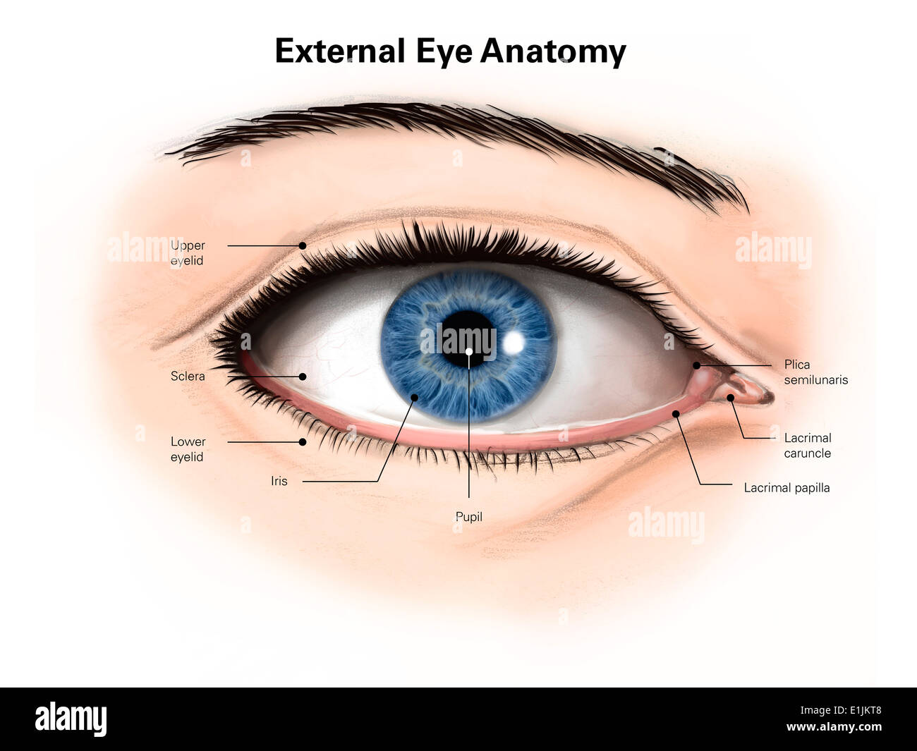
External anatomy of the human eye (with labels Stock Photo: 69866840 - Alamy

Human Eyeball Diagram - YouTube
Post a Comment for "41 human eye with labels"