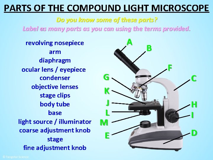
40 microscope parts and labels
microscopeinternational.com › compound-microscopeCompound Microscope Parts, Functions, and Labeled Diagram Nov 18, 2020 · The individual parts of a compound microscope can vary heavily depending on the configuration & applications that the scope is being used for. Common compound microscope parts include: Compound Microscope Definitions for Labels Eyepiece (ocular lens) with or without Pointer: The part that is looked through at the top of the compound microscope ... Microscope Objective Lens | Products | Leica Microsystems Microscope Objectives. Leica Microsystems – The Ultimate in Optical Competence. For more than 170 years Leica Microsystems has designed and produced top-class objectives for a wide variety of applications in research, industry and medicine. The optics specialists at Leica Microsystems bring the highest level of experience and expertise to bear in reducing …
Electron microscope - Wikipedia An electron microscope is a microscope that uses a beam of accelerated electrons as a source of illumination. As the wavelength of an electron can be up to 100,000 times shorter than that of visible light photons, electron microscopes have a higher resolving power than light microscopes and can reveal the structure of smaller objects. A scanning transmission electron microscope …

Microscope parts and labels
en.wikipedia.org › wiki › Scanning_electron_microscopeScanning electron microscope - Wikipedia History. An account of the early history of scanning electron microscopy has been presented by McMullan. Although Max Knoll produced a photo with a 50 mm object-field-width showing channeling contrast by the use of an electron beam scanner, it was Manfred von Ardenne who in 1937 invented a microscope with high resolution by scanning a very small raster with a demagnified and finely focused ... › products › microscopeMicroscope Objective Lens | Products | Leica Microsystems The objective lens is a critical part of the microscope optics. The microscope objective is positioned near the sample, specimen, or object being observed. It has a very important role in imaging, as it forms the first magnified image of the sample. The numerical aperture (NA) of the objective indicates its ability to gather light and largely determines the microscope’s resolution, the ... Label the microscope — Science Learning Hub Jun 08, 2018 · All microscopes share features in common. In this interactive, you can label the different parts of a microscope. Use this with the Microscope parts activity to help students identify and label the main parts of a microscope and then describe their functions.. Drag and drop the text labels onto the microscope diagram. If you want to redo an answer, click on the …
Microscope parts and labels. rsscience.com › stereo-microscopeParts of Stereo Microscope (Dissecting microscope) – labeled ... The objective lenses are the most important parts of a microscope. Compared to a compound microscope where the objectives attached to the nosepiece can be seen and identified individually (based on color bands and their respective labels), the objectives of a dissecting microscope are located in a cylindrical cone and, therefore, are not ... Parts of Stereo Microscope (Dissecting microscope) – labeled … The objective lenses are the most important parts of a microscope. Compared to a compound microscope where the objectives attached to the nosepiece can be seen and identified individually (based on color bands and their respective labels), the objectives of a dissecting microscope are located in a cylindrical cone and, therefore, are not ... › 6-label-the-microscopeLabel the microscope — Science Learning Hub Jun 08, 2018 · All microscopes share features in common. In this interactive, you can label the different parts of a microscope. Use this with the Microscope parts activity to help students identify and label the main parts of a microscope and then describe their functions. Drag and drop the text labels onto the microscope diagram. If you want to redo an ... Scanning electron microscope - Wikipedia A scanning electron microscope (SEM) is a type of electron microscope that produces images of a sample by scanning the surface with a focused beam of electrons.The electrons interact with atoms in the sample, producing various signals that contain information about the surface topography and composition of the sample. The electron beam is scanned in a raster scan …
Microscope Components - Science Quiz - GeoGuessr Microscope Components - Science Quiz: The most common type of modern microscope is called a compound microscope. They have two systems of lenses, one is the eyepiece and the other is comprised of one or more objective lenses. This type of microscope has become so advanced that some are capable of magnifying up to 1000 times! Microscopes are used in … montessorimaterials.org › scienceMontessoriMaterials.org Kingdom Labels Labels used for animal classification ... Wide Parts of Turtle Cards donated by Katie ... Microscope. Microscope ... Wikipedia:Citation needed - Wikipedia To ensure that all Wikipedia content is verifiable, Wikipedia provides a means for anyone to question an uncited claim.If your work has been tagged, please provide a reliable source for the statement, and discuss if needed.. You can add a citation by selecting from the drop-down menu at the top of the editing box.In markup, you can add a citation manually using ref tags. en.wikipedia.org › wiki › Wikipedia:Citation_neededWikipedia:Citation needed - Wikipedia If someone tagged your contributions with a "Citation needed" tag or tags, and you disagree, discuss the matter on the article's talk page.The most constructive thing to do in most cases is probably to supply the reference(s) requested, even if you feel the tags are "overdone" or unnecessary.
MontessoriMaterials.org Kingdom Labels Labels used for animal classification Classification Control Chart. ... Mammals; Myriapod; donated by Colleen; Protozoa; Reptile donated by Katie; Animal Anatomy. Wide Parts of Turtle Cards donated by Katie Turtle Definition Cards donated by Angi Fish: What's Missing?, File 2 ... Microscope. Microscope Nomenclature Cards 1 ... Scientific Method Worksheets - The Biology Corner Microscope Use. How to Use a Microscope – basic guidelines, tips and troubleshooting for the classroom light microscope | Presentation. Microscope Labeling – image, no labels Microscope Coloring – learn the parts of the microscope by coloring. Microscope “E” Lab – use a microscope to examine the letter “e” Compound Microscope Parts, Functions, and Labeled Diagram Nov 18, 2020 · The individual parts of a compound microscope can vary heavily depending on the configuration & applications that the scope is being used for. Common compound microscope parts include: Compound Microscope Definitions for Labels Eyepiece (ocular lens) with or without Pointer: The part that is looked through at the top of the compound microscope ... Label the microscope — Science Learning Hub Jun 08, 2018 · All microscopes share features in common. In this interactive, you can label the different parts of a microscope. Use this with the Microscope parts activity to help students identify and label the main parts of a microscope and then describe their functions.. Drag and drop the text labels onto the microscope diagram. If you want to redo an answer, click on the …
All Saints Online: Diagram for Labelling: Microscope
› products › microscopeMicroscope Objective Lens | Products | Leica Microsystems The objective lens is a critical part of the microscope optics. The microscope objective is positioned near the sample, specimen, or object being observed. It has a very important role in imaging, as it forms the first magnified image of the sample. The numerical aperture (NA) of the objective indicates its ability to gather light and largely determines the microscope’s resolution, the ...

Compound Light Microscope Labeled - Made By Creative Label
en.wikipedia.org › wiki › Scanning_electron_microscopeScanning electron microscope - Wikipedia History. An account of the early history of scanning electron microscopy has been presented by McMullan. Although Max Knoll produced a photo with a 50 mm object-field-width showing channeling contrast by the use of an electron beam scanner, it was Manfred von Ardenne who in 1937 invented a microscope with high resolution by scanning a very small raster with a demagnified and finely focused ...

30 Label Parts Of Microscope - Labels Database 2020
36 Label Parts Of The Microscope - Labels 2021
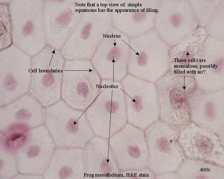
Epithelial Tissue : Anatomy & Physiology

Microscope With Labels Clip Art at Clker.com - vector clip art online, royalty free & public domain
30 Label The Indicated Parts Of The Microscope - Label Ideas 2020

Label And Color The Parts Of Both Microscopes - Best Label Ideas 2019

44+ Label The Parts Of A Microscope Brainly Background – Berita Seputar Dunia Bisnis Terkini
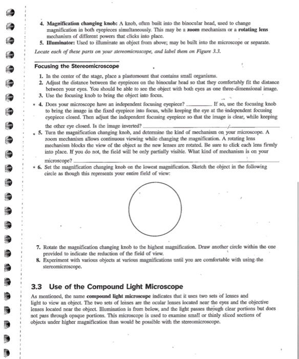
Using the Compound Microscope in Class - Microscopy

Label the Microscope Part

Give the label each part of the microscope: - Brainly.ph

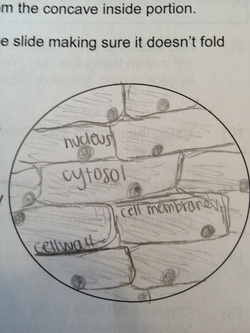
Histology Drawings: January 2014
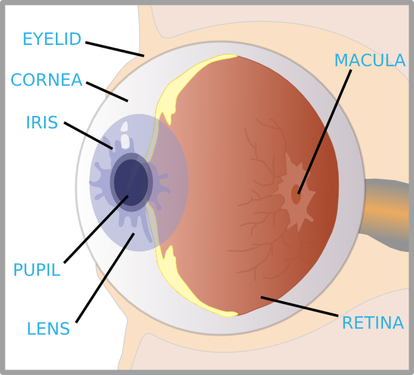
Eye With Labels Clip Art at Clker.com - vector clip art online, royalty free & public domain

Researchers Convert Human Skin Cells into Sensory Neurons - Neuroscience News

microscope labeled microscope worksheet labeling sc 1 st template entrancing labelling - Top ...

Easy Microscope Diagram With Labels - Micropedia

Diagrams of Microscope | 101 Diagrams
Post a Comment for "40 microscope parts and labels"